Showing posts with label Upper Back. Show all posts

Monday, 17 November 2014

Erector Spinae Group

 The Erector Spinae group consists of three back muscles, commonly called 'the Erectors'.
Imagine a tree with 3 branches:

Spinalis
Longissimus
Iliocostalis 

 Together they span the back of your entire spine from the back of your skull (occiput) to the base of your spine (sacurm). Simply put - there's all kinds of attachment points and layers and I don't think I EVER got them memorized in school (!) Well it's time to tackle this group of three - one at at time! But first! As a group (heh) - so yeah! you may want to grab a cup of coffee for this one!


Erector Spinae Group: Attachment Points:

ORIGIN:  

Common tendon (the thoracolumbar fascia) that attaches to the posterior surface of the sacrum, iliac crest, spinous processes of the lumbar vertebrae (L1-L5) and last two thoracic vertebrae (T11-T12)

INSERTION:
Various Attachments at the posterior (back of the) Ribs, spinous & transverse processes of thoracic and cervical vertebrae, and mastoid process of temporal bone.


All together - Erector Spinae group:
Muscle Action(s):
  
UNILATERALLY (one side):

- ipsi-laterally flexes the vertebral column (spine) 
aka side flexion to the same side


side flexion
BILATERALLY (both left & right together)

- extends the spine (spine extension!)

*check out my video "6 movements of the spine" for another visual of side flexion and extension of the spine:


*be sure to checkout "breaking down the spine" to get a better idea of how the spine is divided up - and to get a visual of the transverse processes!


Read on if you dare explore in more detail...

THE BRANCHES! (mwahahahahahaaaaa)

1) Spinalis: 

Spinalis is the smallest of the three erectors and closest to the spine (spine-alis) 
AND can be divided two sections: 

a) Thoracis (the part near the T-spine) 
b) Cervicis (the part near the C-spine).

Together, a & b look like a backwards 'S' (for Ssspinalis!)

ATTACHMENT POINTS for:

a) Thoracis: 
Origin: spinous processes of upper lumbar and lower thoracic vertebrae
Insertion: spinous processes of upper thoracic vertebrae

b) Cervicis:
Origin: ligamentum nuchae, & the spinous process of C7
Insertion: spinous process of the cervicals (except C1!)


2) Longissimus: 
Longissimus is the thickest of the erectors and also the longest (the longgissimest! ;)

So long that IT can be divided into THREE more sections:

a)  Capitis (referring to the head/skull)
b) Cervicis (referring to the neck/cervix)
 c) Thoracis (referring to the thorax/trunk)  
  
ATTACHMENT POINTS for:

 a) Capitis & b) Cervicis:

Origin: transverse processes (TVPS) of upper five thoracic vertebrae (aka T1-T5)
Insertion: a) mastoid process of temporal bone b) TVPS of the the C-spine

 c) Thoracis:

Origin:  Common Tendon (thoracolumbar fascia/aponeurosis)
 Insertion:  lower nine ribs (ribs 4-12) and TVPS of thoracic vertebrae  


3) Iliocostalis: "from hip to rib"
Iliocostalis is the most lateral (furthest away from the spine)
and has long tendons that extend outwards from the hip onto the ribs - underneath the shoulderblades AND can be divided into three sections:

a) Cervicis
b) Thoracis
c) Lumborum

ATTACHMENT POINTS for:

 a) Cervicis & b) Thoracis:
Origin: posterior surface of all the ribs (1-12)
Insertion: a) TVPS of lower cervicals  b) posterior surface of ribs 1-6

 c) Lumborum:

Origin: common tendon (thoracolumbar fascia/aponeurosis)
  Insertion: TVPS of L1-L3 and the posterior surfaces of ribs 6-12


Whewf! Are you still there? We made it you guys!

Monday, 31 March 2014

Rhomboids


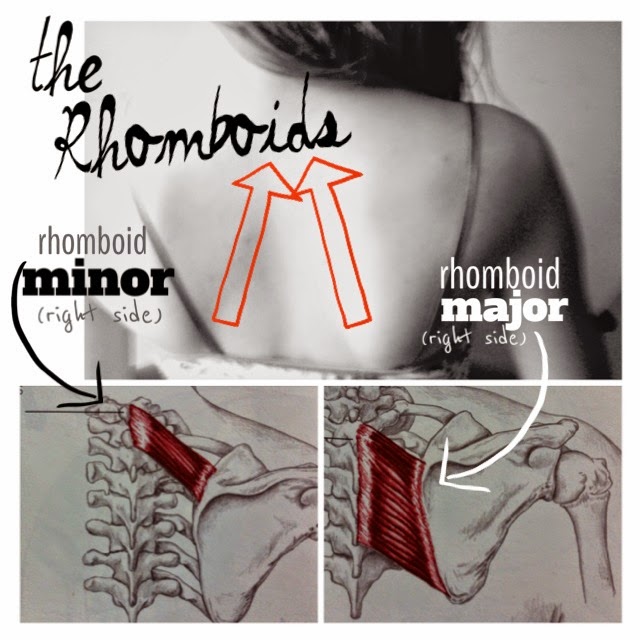
THE RHOMBOIDS:
A PAIR OF MUSCLES: Major & Minor (BFFs!)

These upper back muscles sit on an angle between your spine and your shoulder blade!* There is RHOMBOID MAJOR and just above (and about half its size) is RHOMBOID MINOR:
* More specifically:

RHOMBOID MAJOR attaches to the 2nd through 5th thoracic vertebrae (T2-T5) > crossing at a downward angle and attaching to the MEDIAL (inside) border of the SCAPULA (shoulder blade)

**even more specifically > between the root of the spine of the scapula & the inferior angle of the scapula**

*RHOMBOID MINOR sits above Major > attaching to vertebrae (spine) C7-T1 
> C7 is the last vertebrae of neck bones > & T1 is the first vertebrae of the thoracic spine.

RHOMBOID MUSCLE ACTIONS:

 When contracted, both Rhomboid Major & Minor work together to perform THREE different muscle actions.  All actions are moving the SCAPULA (shoulderblade) at the SCAPULOTHORACIC JOINTS (across the back of the ribcage) aka S/T joint
(but can also be referred to as S/C joints >aka SCAPULO-COSTAL joints (costal meaning ribs))


The 3 actions are:

1-RETRACTION of the scapula at the s/t joints (shoulderblades intending towards the spine (centerline of body) >causing chest to open)

2-ELEVATION of the scapula at the s/t joints (this looks like a shoulder-shrug)

3-DOWNWARD ROTATION of the scapula at the s/t joints (pointed corner of shoulderblade (looks like an upside-down triangle) swings down and in toward the spine (centerline of body))


Most of us are battle posture issues like rounded shoulders and head forward, the more we hunch over - the more this encourages muscle imbalance.  Chest muscles get tight, we round forward causing our shoulder blade to sit further and further away from each other!  Poor RHOMBOIDS get over-stretched, contracting becomes difficult and rhomboids become weak.
Here is an easy way to fight this muscle imbalance! And all you need is a wall!

SCAPULAR WALL SLIDES:

- begin by squatting slightly with your back against the wall, heels of feet are not touching the wall > knees softly bent, knees & ankles aligned (without knee bending forward past ankle!) What IS touching the wall is your bum and upper back.

-then place backs of your arms against the wall in a 90/90 degree position which looks like the "don't shoot!" position - see picture :) this is the FIRST position

- next you will slowly raise your arms together whilst keeping your elbows and backs of hands flat against the wall.(they may want to 'lift off' you'll see ;)

-Remember to contract your back muscles at the same time, intending shoulder blades to touch (intending, not forcing)
MOVE SLOWLY to avoid injury. Know your limits and trust your intuition.  If you feel shaking that's okay it means your rhomboids need this! If its difficult then your rhomboids probably need this! If discomfort crosses that line into pain/the threat of injury) slowly lower arms and back out.

Repeat this up and down motion nice and slow - like!  For strengthening I like to recommend 10 reps x 3 sets or until muscles feel exhausted. 

DO THIS EVERY DAY TO ENCOURAGE A happy and balanced posture! *note there will most likely be a sore muscle/transition period if these muscles are not used to working so much ;) warm epsom salt baths. rest and drinking lots of water will help speed along this muscle soreness ;)

I love showing this one to my clients because I feel it doubles as a chest opener as well! - time management!


Show your Rhomboids some love today!

Monday, 3 March 2014

Splenius Cervicis & Splenius Capitis


Splenius Cervicus & Splenius Capitis = BFFs <3

Both muscles run along the back of your neck and spine. I used to think our neck muscles ended when our shoulders and back began but nope! 
(Something to consider when you ask for a neck massage!)
Splenius Cervicis starts up at the very top vertebrae in your neck (c1-c3) and reaches down to the thoracic vertebrae (T3-T6 -between shoulder blades)

Splenius Capitis has all kinds of attachment points : starting at the bone just behind your ear & the back of your skull (aka occiput), down the back of the neck c3-c6 , continuing down from c-spine into the thoracic spine (c7 -T4) !! Major!

Cervicis: think neck (aka cervical spine)
Capitis : think head (because it starts on the skull)

Each of these Splenius neck muscles
Form a 'V shape' !

MUSCLE ACTIONS:

They both perform practically the same actions !
Splenius Capitis and Splenius Cervicis both do the same 3 actions: here's a visual! 

1. Ipsi-lateral flexion (aka- ear to shoulder on the side the muscle is on)

2. Ipsi-lateral rotation (aka- turning head to same side muscle is on) 

3. Head and neck extension (aka-looking up!) 

The main movement of the Splenii is neck extension - these muscles are often referred to as "the neck extensors" and are often tight on most people I treat due to poor posture (head forward position) most common seen in computer/desk workers!